Immersion at the SOLEIL synchrotron: a major scientific facility for exploring matter
Immersion at the SOLEIL synchrotron: a major scientific facility for exploring matter
How can we examine the structure of biomass without destroying it? How can we identify the chemical elements present in a sample or reveal structures that are invisible to conventional laboratory instruments? To answer these questions, researchers can draw on an exceptional tool: the SOLEIL synchrotron.
During the CHEMIMAG 2026 scientific conference dedicated to multi-scale chemical imaging, organised on 3 July by the PEPR B-BEST, participants were able to visit the SOLEIL synchrotron. Here’s a look at this unique scientific instrument, how it works and the opportunities it offers to researchers.
1. The SOLEIL synchrotron: a ‘super-lamp’ for exploring matter
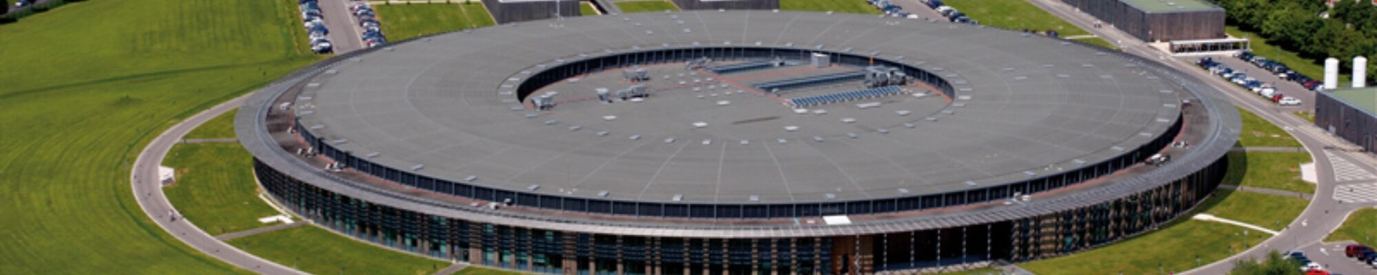
Located on the Saclay plateau, the SOLEIL synchrotron – the optimised intermediate-energy light source of the LURE (Laboratory for the Use of Electromagnetic Radiation) – is a major French scientific instrument, funded by the CNRS and the CEA. It works by producing extremely intense light, known as synchrotron radiation, which enables matter to be explored with remarkable precision.
This light covers a very broad spectral range, from infrared to X-rays, thus offering a wide array of analytical techniques. Highly stable and with minimal dispersion, it provides a resolution far superior to that of conventional laboratory equipment.
Thanks to these capabilities, the synchrotron enables the analysis of a wide variety of samples, ranging from entire organisms down to the atomic scale. It is used in numerous disciplines: biology, medicine, geosciences, chemistry, materials science, agri-food, astrophysics, palaeontology and the study of cultural heritage.
2. How does the SOLEIL synchrotron work?
A synchrotron is an electromagnetic instrument designed to accelerate electrons in order to produce powerful light known as synchrotron radiation.
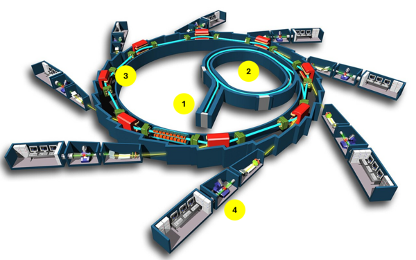
It all begins in an electron gun. A tungsten target, roughly the size of a two-euro coin, is heated by an electric current. This energy causes electrons to be emitted from the metal. This is known as thermionic emission. A gold grid, situated opposite the tungsten pellet, then creates a potential difference that attracts and accelerates these electrons.
Step 2: accelerating the electrons
The electrons enter a linear accelerator where they reach a speed close to that of light before being injected into a first circular accelerator, known as a booster. In this circular accelerator, the electrons gain even more speed thanks to bending magnets that guide their trajectory and radio-frequency cavities that restore their energy.
Step 3: producing light
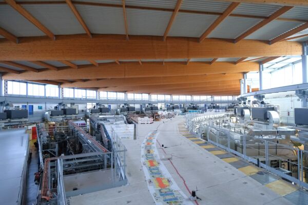
The electrons are then sent into a storage ring shaped like a polygon, where they circulate continuously. At each change of direction within the ring, the electrons generate a beam of light through energy loss: this is known as synchrotron radiation.
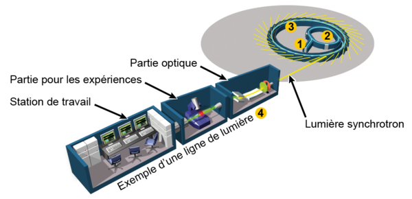
This light is then directed towards 29 beamlines installed around the ring. SOLEIL’s beamlines are the experimental facilities used to study the samples. Each beamline utilises a specific part of the synchrotron radiation spectrum and employs specific analytical techniques to study the structure and properties of the material samples.
3. Facilities open to the entire scientific community
The SOLEIL synchrotron is accessible to the French and international scientific communities, as well as to industry, via calls for proposals. Scientists whose institutions are based in France benefit from free access to the facilities, as well as accommodation and meals for three people per project.
A call for proposals is currently open and closes on 15 September 2026, for use from early March 2027 to late July 2027: view the call for proposals.
In addition to the equipment, the SOLEIL synchrotron provides the expertise of its scientists, who support teams at every stage, from sample preparation and the choice of techniques to the analysis of results.
4. An example of use: the FillingGaps project
Among the projects in the Bioproductions research programme, the FillingGaps project utilised the SOLEIL synchrotron to investigate the enzymatic degradation of plant biomass at different scales. Two beamlines were used: DISCO and ANATOMIX, whose complementary approaches make it possible to link the chemical composition of plant cell walls to their structural organisation.
On the DISCO beamline, deep-ultraviolet (Deep-UV) fluorescence microscopy experiments were carried out on various samples from the project, including poplar wood, maize stalks and algae. One of the main advantages of this beamline is access to the deep-UV spectral range for microscopy, which is inaccessible to laboratory microscopes and exclusive to the SOLEIL synchrotron. This excitation makes it possible to reveal the autofluorescence of phenolic molecules, such as lignin and hydroxycinnamic acids, which are involved in the resistance of plant cell walls to enzymatic degradation. It is thus possible to map their location, estimate their relative abundance and track changes in their signal during degradation.
One of the flagship experiments conducted on DISCO involves monitoring the enzymatic degradation of plant cell walls in real time. This approach simultaneously highlights the autofluorescence of the phenolic compounds in the cell walls and that of the enzymes themselves, which is linked to the presence of aromatic amino acids that fluoresce in the deep UV range. This unique capability makes it possible to visualise the interactions between enzymes and their substrates, to study the dynamics of degradation and, in conjunction with other analytical techniques, to gain a better understanding of the mechanisms responsible for biomass recalcitrance.
As these observations are made on thin sections, they only provide a local picture of the heterogeneity of the samples. In order to study structural changes on a more representative scale, X-ray tomography experiments were carried out on the ANATOMIX beamline. Thanks to synchrotron radiation, this technique offers a spatial resolution far superior to that of laboratory tomographs, with a voxel size of 0.65 µm. It enables the three-dimensional visualisation of anatomical changes induced by enzymatic degradation and allows these to be correlated with observations obtained using UV microscopy.
Thanks to Amandine LEROY, Postdoctoral Researcher at DISCO Synchrotron SOLEIL (ST-AUBIN), for her contribution to the drafting of this article.
By browsing our site you accept the installation and use cookies on your computer.
Know more
Our use of cookies
Cookies are a set of data stored on a user’s device when the user browses a web site. The data is in a file containing an ID number, the name of the server which deposited it and, in some cases, an expiry date. We use cookies to record information about your visit, language of preference, and other parameters on the site in order to optimise your next visit and make the site even more useful to you.
To improve your experience, we use cookies to store certain browsing information and provide secure navigation, and to collect statistics with a view to improve the site’s features. For a complete list of the cookies we use, download “Ghostery”, a free plug-in for browsers which can detect, and, in some cases, block cookies.
You can also visit the CNIL web site for instructions on how to configure your browser to manage cookie storage on your device.
In the case of third-party advertising cookies, you can also visit the following site: https://www.youronlinechoices.com/fr/controler-ses-cookies/, offered by digital advertising professionals within the European Digital Advertising Alliance (EDAA). From the site, you can deny or accept the cookies used by advertising professionals who are members.
It is also possible to block certain third-party cookies directly via publishers:
Cookie type
Means of blocking
Analytical and performance cookies
Realytics Google Analytics Spoteffects Optimizely
Targeted advertising cookies
DoubleClick Mediarithmics
The following types of cookies may be used on our websites:
Mandatory cookies
Functional cookies
Social media and advertising cookies
These cookies are needed to ensure the proper functioning of the site and cannot be disabled. They help ensure a secure connection and the basic availability of our website.
These cookies allow us to analyse site use in order to measure and optimise performance. They allow us to store your sign-in information and display the different components of our website in a more coherent way.
These cookies are used by advertising agencies such as Google and by social media sites such as LinkedIn and Facebook. Among other things, they allow pages to be shared on social media, the posting of comments, and the publication (on our site or elsewhere) of ads that reflect your centres of interest.
Our EZPublish content management system (CMS) uses CAS and PHP session cookies and the New Relic cookie for monitoring purposes (IP, response times). These cookies are deleted at the end of the browsing session (when you log off or close your browser window)
Our EZPublish content management system (CMS) uses the XiTi cookie to measure traffic. Our service provider is AT Internet. This company stores data (IPs, date and time of access, length of the visit and pages viewed) for six months
Our EZPublish content management system (CMS) does not use this type of cookie.
For more information about the cookies we use, contact INRAE’s Data Protection Officer by email at cil-dpo@inra.fr or by post at:
INRAE 24, chemin de Borde Rouge –Auzeville – CS52627 31326 Castanet Tolosan cedex - France